
Aneurysmal bone Cyst
Clinical History:
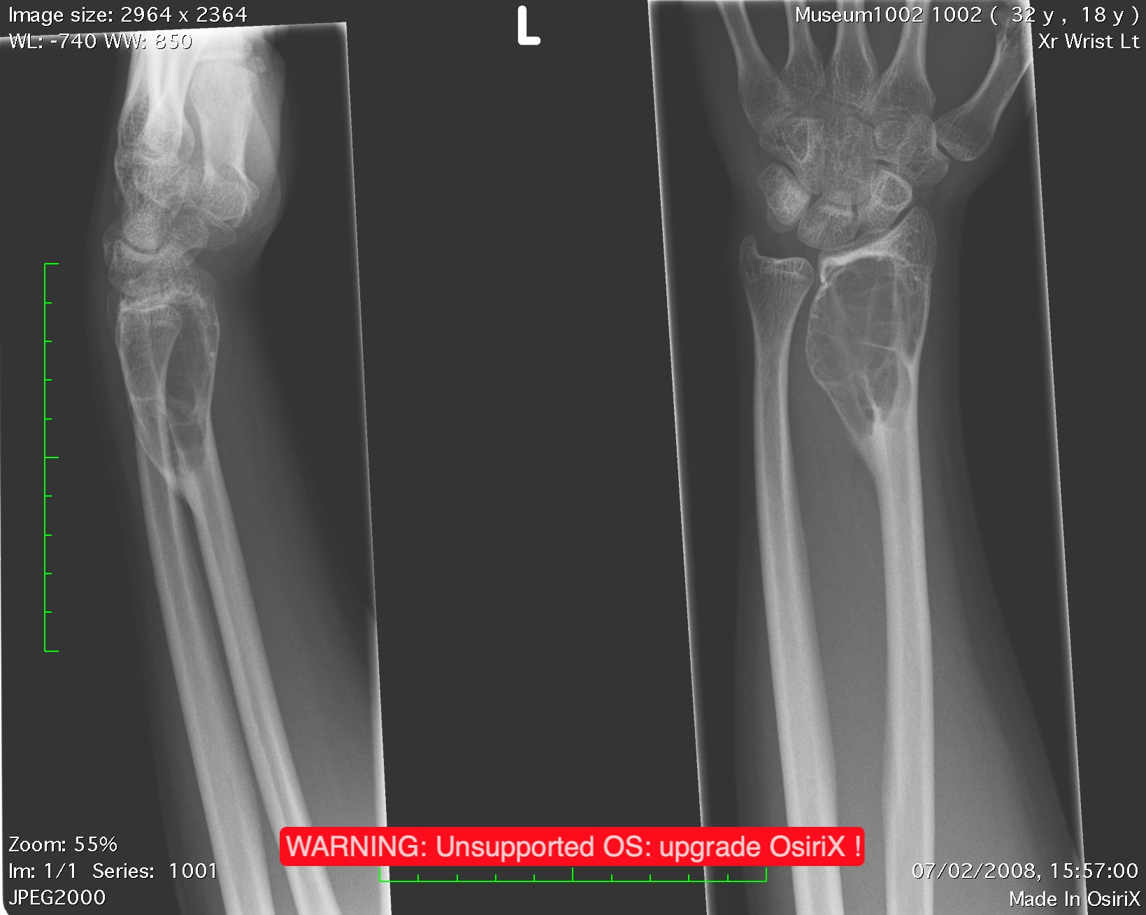
• Pain left wrist. No history of trauma.
Radiological Findings:
- The plain film demonstrates a lytic lesion within the metaphysis and epiphysis reaching a subarticular position.
There is an expansion and thinning of the cortex. No cortical breach. No soft tissue mass. No identifiable matrix calcification or ossification.
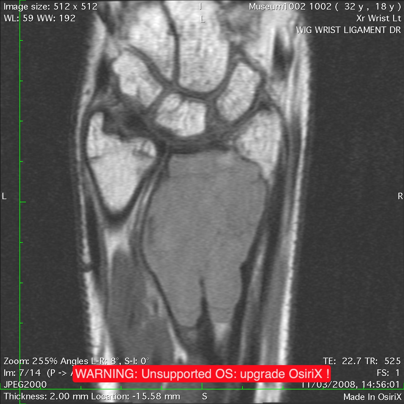
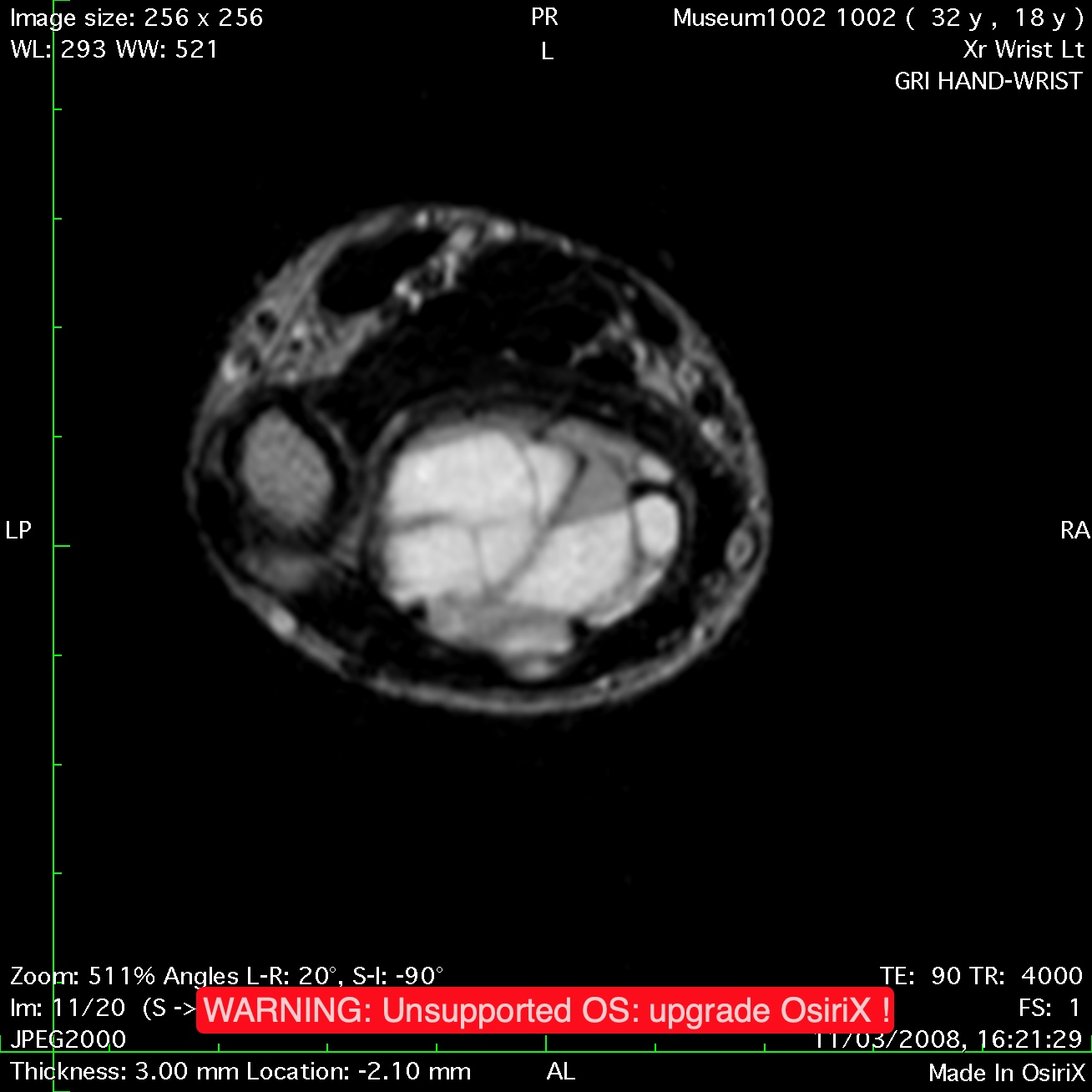
The MRI shows the lesion to be confined within the bony structures.
There is evidence of fluid fluid levels.
No soft tissue mass.
Differential Diagnosis:
Giant cell tumour.
Aneurysmal bone cyst.
Fibrous dysplasia.
Principal Diagnosis:
Aneurysmal bone cyst.
Additional Information:
• This is a slightly unusual diagnosis in a 21-year-old. An aneurysmal bone cyst is usually seen before epiphyseal fusion. It reaches a subarticular position and is characterized by this. Fluid-fluid levels on MRI imaging are highly suspicious but not diagnostic of an aneurysmal bone cyst. A giant cell tumour is a good alternative diagnosis particularly in a patient of this age. Subarticular lesions in a 21-year-old would more commonly be due to giant cell tumours.