
ESSR Protocols for Elbow MRI

ESSR guidelines for Elbow MRI Planes
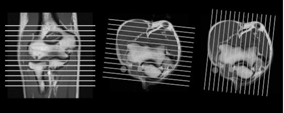
What to look in each plane?
|
Axial:
|
 |
|
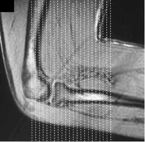
Sagittal:
|
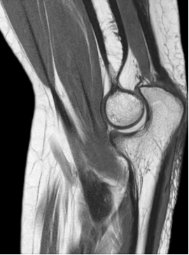 |
|
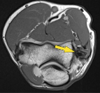
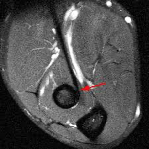
Coronal:
|
(Courtesy: Supermedica.com) |
|
FABS:
|
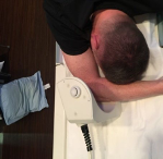 |
 |
|
 |

What to look in each sequenc?
SEQUENCES |
USES:
|
T1W |
|
PDW |
|
Fat Suppressed T2 or PD |
|
T2* weighted |
|
Arthrograms |
|
Elbow MRI Checklist
Bone |
Soft Tissues |
|
|
Elbow joint |
Nerves |
|
|
Standard MRI Report
|
D. STANDARD REPORTS
|
FINDINGS:
No evidence of joint effusion.
Normal signal intensity of the bone marrow with no focal lesions.
Intact juxta-articular muscles and tendons.
No features of epicondylitis.
Normal proximal radioulnar articulation.
Intact ulnar and radial collateral ligaments.
No definite fracture noted.
Normal signal and thickness of the ulnar nerve.
CONCLUSION:
Normal MRI of the elbow with no evidence of fluid collection.