ESSR planes for Hip MRI

Text & media

What each plane is used for?
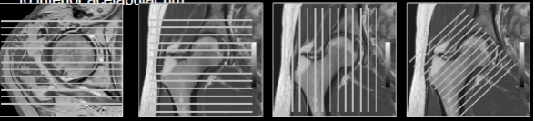
Axial:

  • Acetabular version
  • Cam lesion (FAI)
  • Labrum.
  • Iliopsoas Muscles.
  • Gluteal Muscles.
  • Proximal femoral version.
  • Paralabral cysts.
  • Collections.
  • Ischiofemoral space
  • Hamstring tendons


127

127

(Courtesy:www.minclinic.ru)


Sagittal:

  • Cartilage assessment
  • Bone assessment for OA
  • Iliopsoas tendonitis/bursitis
  • Anterosuperior labrum


125


Coronal:

  • Femoral Head congruency/subchondral fracture/cyst.
  • Joint fluid assessment.
  • Gluteal tendon insertion.


126


Axial Oblique

  • For cam deformity of femur


128


129

(Courtesy: www.radsource.us)


Coronal Oblique

  • Isolated hip assessment


129


129

(Courtesy: clinicalgate.com)

Text & media

SEQUENCES

USES:


T1W 

  • Anatomical Details.
  • Marrow signal
    1. Fractures
    2. AVN
    3. Tumours

PDW

  • Anatomical details.
  • Cartilage assessment.


Fat Suppressed T2 or PD

  • Gluteal Tendinopathy
  • Tendon tears.
  • Bone Bruising
  • Muscle oedema 
  • (Q.Femoris in Ischiofemoral impingement)
  • Effusion. 
  • Labral tears 
  • Para labral cysts


Arthrograms

  • Labral tears
  • FAI 
  • Cartilage Assessment

Text & media

Bone and Joint.

Soft Tissues

  • AVN Femoral head – mid-age female.
  • Fracture NOF - elderly
  • Joint effusion
  • Osteomyelitis
  • Septic arthritis
  • Cares Sicca – Dry arthritis TB
  • OA / Inflam. arthropathy
  • Mets (Subtrochanteric)
  • ASIS, AIIS Avulsion–athletes
  • Adductor avulsion
  • Hamstring avulsion
  • FAI and Labral tears
  • DDH
  • Sacroiliitis
  • Transient osteoporosis
  • Cares Sicca – Dry arthritis TB
  • Osteoid Osteoma
  • Stress fractures
  • Rarely Primary bone tumours


  • Gluteus Medius tendonitis
  • Gluteus Medius tendon tear
  • Trochanteric bursitis.
  • Morel Lavallee’ lesion.
  • Hamstring avulsion tear
  • Myositis.
  • Muscle tear.
  • Groin hernia.
  • Ischiofemoral impingement
  • Avulsion ligament head of femur.
  • Rarely Other tendon tears including-
  • Hamstring avulsion
  • Adductor longus tear


Standard MR Report

And diagnostic Algorithm


D. STANDARD REPORTS


MRI HIP


FINDINGS:

  • Preserved hip joint spaces bilaterally. Preserved chondral surfaces.
  • No fracture or dislocation. Normal femoral head coverage.
  • No evidence of avascular necrosis.
  • Unremarkable sacroiliac joints.
  • Unremarkable periarticular muscles and soft tissues.
  • Normal abductor tendons. No trochanteric bursitis.
  • No focal marrow abnormality.

IMPRESSION:

Normal MRI of the hip joints.
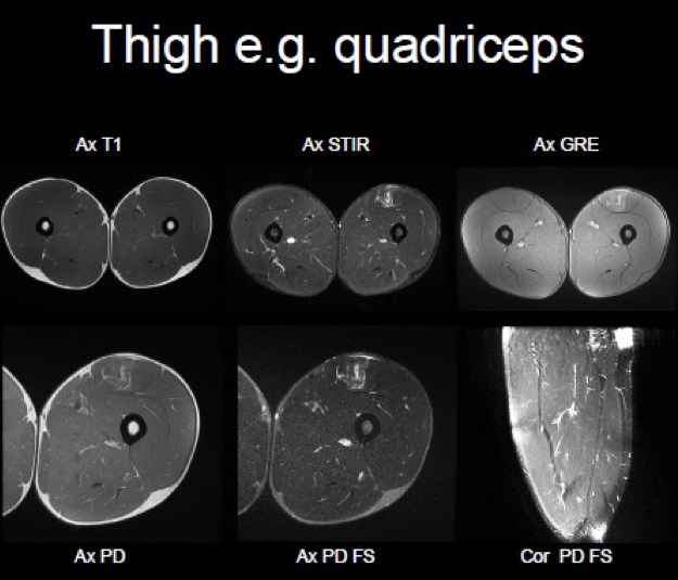


MRI THIGH


FINDINGS:

  • Normal left hip joint and left femoral head with no evidence of AVN.
  • Preserved muscle bulk and architecture with no focal tear.
  • No myofascial or musculotendinous junction oedema.
  • No focal soft tissue abnormality.
  • Normal neurovascular bundle.
  • No focal marrow lesions.


CONCLUSION:

Unremarkable MRI study of the thigh.


E. APPLIED RADIOLOGY


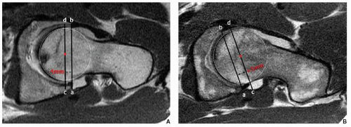
1. Femoro-acetabular impingement (FAI):

The surgical treatment will change with the cause and effects of FAI.

  • Cam deformity can be treated with shaving the ‘bump’ at the lateral aspect of the femoro-acetabular junction.
  • If there is associated labral tear, then shaving the ‘bump’ + labral repair.
  • If there is chondral changes or degeneration then may require shaving the patient may require resurfacing of the hip joint.


2. Always check for the SI joints.


3. In thigh MRI trauma cases, always check for rectus femoris central aponeurosis.