
ESSR Planning images for Knee MRI
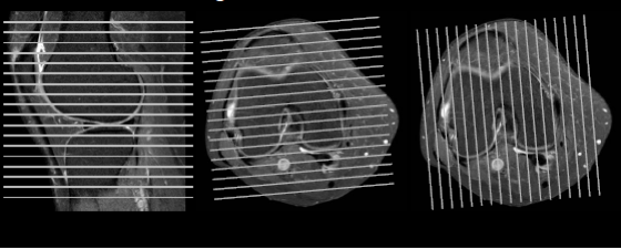
Image Sequences

Use of Each Imaging Plane
|
Sagittal:
|
|
|
Axial:
|
|
|
Coronal:
|
|


|
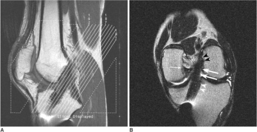
Coronal Oblique ACL assessment.
Ref: Grading anterior cruciate ligament graft injury after ligament reconstruction surgery: diagnostic efficacy of oblique coronal MR imaging of the knee. Moon SG, Hong SH, Choi JY, Jun WS, Choi JA, Park EA, Kang HS, Kwon JW - Korean J Radiol (2008 Mar-Apr)
|
|

Checklist
Menisci /Cartliage |
Cruciate ligaments |
|
|
Collateral ligaments |
Patellofemoral Joint |
|
|
Various |
Patellar tendon |
|
|
Text & media
|
D. STANDARD REPORTS
|
FINDINGS:
- No joint effusion.
- Preserved patellofemoral alignment with no trochlear dysplasia. Intact patellofemoral ligaments. Normal extensor mechanism.
- No meniscal tear seen. Intact cruciate ligaments.
- Preserved chondral surfaces.
- Normal posterolateral and posteromedial corner structures.
- Intact collateral ligaments.
- No focal bone lesions.
- No popliteal fossa abnormality.
IMPRESSION:
Small joint effusion, otherwise unremarkable.
Intact menisci.
Intact cruciate ligaments.
REPORT 2:
FINDINGS:
- Horizontal tear seen in the posterior horn of the lateral meniscus.
- Intact medial meniscus.
- Moderate joint effusion is present.
- Intact medial and lateral collateral ligaments.
- There is no bony injury.
- Intact anterior and posterior cruciate ligaments.
- IMPRESSION:
Lateral meniscus tear.
Joint effusion.
|
E. APPLIED RADIOLOGY
|
ACL Tear
Check Bruising pattern
Check Posterolateral corner injury
Meniscal injury
Sulcus sign
Tibial translation
ACL graft – Check
Loosening anchors in tunnels
Check tibial tunnel angle and Blumensaat’s line
Graft patency/ laxity
Cyclops lesion
Meniscal tear
Parameniscal cyst
Bucket handle/ flipped fragment/ flap tear
Meniscal root tear
OCD
Size of lesion
Stability
Loose bodies
Marrow oedema
Subchondral insufficiency/ stress fracture